Dr Alan Cheung
Senior Consultant Orthopaedic Surgeon in Singapore
MBBS (London), MRCS (England), FRCS (Trauma and Orthopaedics, England), Diploma in Sport and Exercise Medicine (UK), Fellow of the European Board of Orthopaedics and Traumatology, American Academy of Orthopaedic Surgeons (AAOS)
MRI reports are commonly used in meniscus tear diagnosis because they provide detailed images of the meniscus and other structures within the knee. However, radiology reports often contain technical terms such as "meniscal extrusion", "grade 3 signal" and "bucket-handle tear" that may be unfamiliar to patients. Understanding these findings can provide greater clarity about the nature of a meniscus injury and its potential impact on knee function.
Why MRI Is Important for Meniscus Tear Diagnosis
The meniscus is a C-shaped piece of cartilage that acts as a cushion between the thigh bone and shin bone. Each knee has two menisci: the medial meniscus on the inner side of the knee and the lateral meniscus on the outer side.
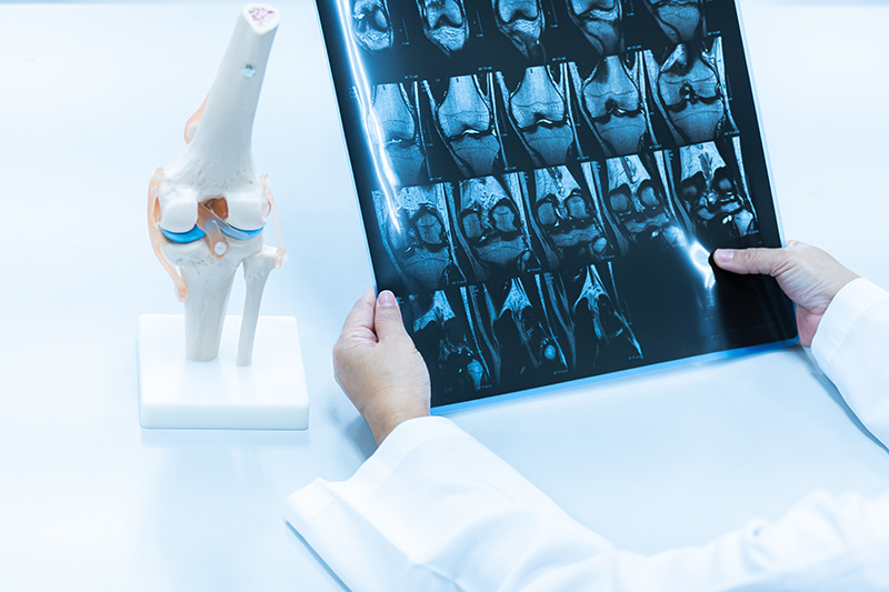
When a meniscus tear is suspected, an MRI is often recommended because it provides detailed images of the soft tissues within the knee. Unlike X-rays, which primarily show bones, MRI scans can reveal damage to cartilage, ligaments and other structures.
MRI findings are only one part of the diagnostic process. A thorough meniscus injury examination, including a review of symptoms and physical assessment, helps doctors determine the extent of the injury and the most appropriate treatment plan.
Understanding Common Terms in a Meniscus MRI Report
Medial Meniscus vs Lateral Meniscus
These terms refer to the two menisci in the knee. The medial meniscus is located on the inner side of the joint and is more commonly injured due to its reduced mobility. The lateral meniscus sits on the outer side and is generally more flexible.
Meniscal Degeneration
Meniscal degeneration describes gradual wear and tear within the cartilage. It is commonly seen as people age and does not always indicate a tear. Degeneration may occur over many years and can sometimes contribute to knee pain or stiffness.
Grade 1, 2 and 3 Signals
Radiologists often use grading systems to describe changes within the meniscus:
- Grade 1 signal: Mild changes within the cartilage without a tear.
- Grade 2 signal: More noticeable internal changes but still not considered a true tear.
- Grade 3 signal: A signal that extends to the surface of the meniscus and is generally considered indicative of a meniscal tear.
These grades help doctors interpret MRI findings as part of a meniscus injury test and determine whether treatment is needed.
Meniscal Tear
A meniscal tear occurs when the cartilage develops a crack or split. Tears can result from sports injuries, sudden twisting movements or age-related degeneration. The size, location and pattern of the tear can affect symptoms and treatment options.
Meniscal Extrusion
Meniscal extrusion means that part of the meniscus has shifted outward from its normal position. This can reduce the meniscus's ability to absorb shock and may be associated with arthritis or more severe tears.
Complex Tear
A complex tear involves multiple tear patterns occurring together. Because the damage affects the meniscus in several directions, these tears can be more challenging to treat than simpler tear types.
What Different Types of Meniscus Tears Mean
MRI reports often specify the pattern of a tear because different tear types can influence treatment recommendations and recovery outcomes.
Horizontal Tear
A horizontal tear runs parallel to the surface of the meniscus, dividing it into upper and lower sections. These tears are commonly associated with age-related degeneration.
Radial Tear
A radial tear begins at the inner edge of the meniscus and extends outward. Because it disrupts the normal structure of the cartilage, it can significantly affect the meniscus's ability to distribute weight across the knee.
Longitudinal Tear
A longitudinal tear runs along the length of the meniscus. Depending on its size and location, this type of tear may be suitable for repair rather than removal.
Bucket-Handle Tear
A bucket-handle tear is a large longitudinal tear in which a portion of the meniscus becomes displaced. This can cause symptoms such as knee locking, restricted movement and significant discomfort.
Flap Tear
A flap tear occurs when a fragment of cartilage becomes partially detached, creating a flap-like section. This loose tissue may catch within the joint and contribute to pain or mechanical symptoms.
Root Tear
The meniscal root anchors the meniscus to the bone. A root tear affects this attachment point and can severely reduce the meniscus's ability to absorb forces within the knee. These injuries often require specialist assessment.
Other MRI Findings That May Accompany a Meniscus Tear
MRI reports often identify additional findings beyond the meniscus. These findings may help explain symptoms and influence treatment decisions.
Bone Marrow Oedema
Bone marrow oedema refers to swelling within the bone, often described as a bone bruise. It may occur following trauma and can contribute to pain during weight-bearing activities.
Joint Effusion
Joint effusion is the accumulation of excess fluid within the knee joint. It is a common response to injury and inflammation and may cause visible swelling.
Cartilage Damage
The MRI may identify areas where the smooth cartilage covering the bones has become worn or damaged. Cartilage injuries can contribute to pain, stiffness and the development of osteoarthritis.
ACL Injury
The anterior cruciate ligament (ACL) is one of the major stabilising ligaments in the knee. Meniscus tears frequently occur alongside ACL injuries, particularly in sports-related accidents involving twisting movements.
Baker's Cyst
A baker's cyst is a fluid-filled swelling that develops behind the knee. It often forms when excess joint fluid accumulates due to conditions such as a meniscus tear or arthritis.
Discuss Your MRI Results With Dr Alan Cheung
MRI reports can provide valuable information about the location, severity and characteristics of a meniscus injury. Understanding commonly used terms such as meniscal degeneration, meniscal extrusion and different tear patterns can help you better interpret your MRI findings and participate more actively in discussions about your care. However, MRI results should always be considered alongside your symptoms, medical history and physical examination findings to ensure an accurate diagnosis and appropriate treatment plan.
At International Orthopaedic Clinic, we are committed to helping patients understand their condition and make informed decisions about their knee health. Under the care of Dr Alan Cheung, patients who have undergone an MRI scan or sustained a meniscus injury can receive a comprehensive assessment of their findings, along with personalised treatment recommendations tailored to their symptoms, activity levels and recovery goals.
If you have recently undergone a meniscus injury test or received an MRI report that you would like explained, contact us today to discuss your results with an orthopaedic specialist.
FAQs About Meniscus Tear Diagnosis and MRI Findings
Does a meniscus tear always appear clearly on an MRI report?
Not always. While MRI is highly accurate for detecting meniscus injuries, some tears may be subtle or difficult to identify depending on their size, location and imaging quality. This is why MRI findings are interpreted together with the results of a meniscus injury examination.
Why does my MRI report mention a meniscus tear if I do not have knee pain?
MRI scans can sometimes detect meniscal changes that are not causing symptoms. In some cases, age-related degeneration or small tears may be present without affecting knee function. Your doctor will determine whether the MRI findings are clinically significant.
Why do MRI reports sometimes describe both a tear and degeneration?
Degeneration and tears can occur at the same time. As the meniscus naturally wears down with age, it can become more vulnerable to injury. This means an MRI report may show signs of both wear and tear and a meniscus tear in the same knee.