


Dr Alan Cheung
Senior Consultant Orthopaedic Surgeon in Singapore
MBBS (London), MRCS (England), FRCS (Trauma and Orthopaedics, England), Diploma in Sport and Exercise Medicine (UK), Fellow of the European Board of Orthopaedics and Traumatology, American Academy of Orthopaedic Surgeons (AAOS)
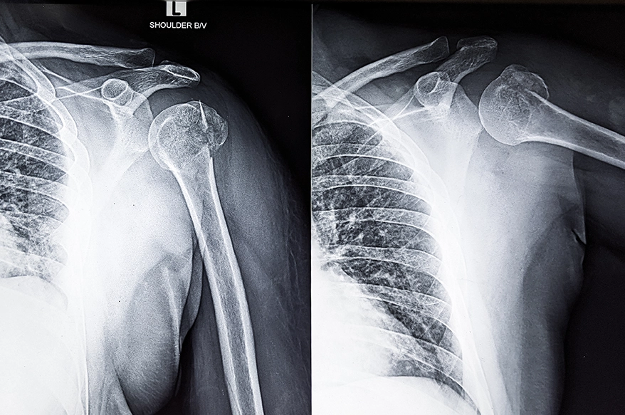
Shoulder dislocations are classified according to the direction in which the upper arm bone moves out of the shoulder socket. Anterior dislocations are the most common, while posterior and inferior dislocations occur less frequently.
The direction can provide clues about how the injury occurred and which surrounding structures may also have been damaged. Together with the patient’s age, activity demands and associated injuries, these findings help guide treatment and rehabilitation.
What Are the Different Types of Shoulder Dislocation?
A shoulder dislocation occurs when the ball of the upper arm bone (humeral head) moves out of the shoulder socket (glenoid). The type of shoulder dislocation is classified according to the direction in which the humeral head has moved. Identifying the direction of the dislocation is an important part of diagnosis, as different directions can be associated with different injury mechanisms and patterns of damage.
Anterior Shoulder Dislocation
Anterior shoulder dislocation is the most common type, accounting for the vast majority of shoulder dislocations. It occurs when the humeral head moves forwards out of the socket, usually after a fall onto an outstretched arm or a forceful impact while the arm is abducted and externally rotated.
It may also damage the labrum, ligaments or rotator cuff. Recurrent instability is an important consideration after an anterior dislocation, particularly in younger and more active patients.
Posterior Shoulder Dislocation
Posterior shoulder dislocation occurs when the humeral head is displaced behind the shoulder socket. It is much less common than anterior dislocation and is often associated with seizures or electrical injuries, although it can also occur after trauma.
Because the symptoms can be subtle and the injury may not always be obvious on initial X-rays, posterior shoulder dislocations are sometimes diagnosed later than anterior dislocations. Early recognition helps prevent ongoing pain, stiffness and shoulder dysfunction.
Inferior Shoulder Dislocation
Inferior shoulder dislocation is a rare injury in which the humeral head moves below the shoulder socket. Patients often present with the arm fixed above the head and are unable to lower it comfortably.
This injury usually results from high-energy trauma and may occur alongside nerve, blood vessel or soft tissue injuries. Prompt specialist assessment is important to evaluate the extent of damage and guide treatment.
Other Rare Shoulder Dislocations
Other directions of glenohumeral dislocation are exceptionally rare and are generally associated with significant trauma and accompanying injuries. These unusual injury patterns require careful assessment and imaging to determine the extent of damage and appropriate treatment.
Why Does the Direction of a Shoulder Dislocation Affect Diagnosis?
The direction of displacement can provide clues about the mechanism of injury and the structures that may have been affected. This becomes particularly important when the injury is less obvious clinically or associated damage is suspected.
Identifying Associated Injuries
Each type of shoulder dislocation is associated with different patterns of injury. Depending on the direction of the dislocation, there may be damage to surrounding soft tissues or bone. Nerves and blood vessels may also be affected in more severe injuries. Identifying these associated injuries helps determine whether further imaging or surgical treatment may be required.
Choosing the Right Imaging Tests
Although X-rays are usually the first imaging investigation, they may not always provide a complete picture, particularly for less common dislocations. Posterior shoulder dislocations, for example, can sometimes be overlooked on standard X-ray views. Additional imaging such as CT or MRI may be recommended when more detail is needed about fractures, bone loss or associated soft-tissue injuries that could influence treatment.
How Does the Direction of a Shoulder Dislocation Influence Treatment and Recovery?
Although the primary goal of treatment is to safely relocate the shoulder joint, the direction of the dislocation influences many aspects of management. The direction of the dislocation can influence the reduction technique and indicate which associated injuries should be considered. Treatment and recovery also depend on the extent of tissue or bone damage and the patient’s risk of recurrent instability.
Initial Management
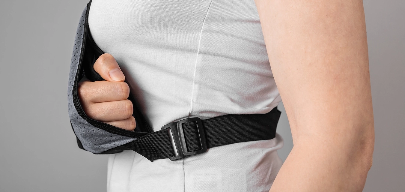
The first priority is to return the humeral head to its normal position, a procedure known as reduction. The technique used may differ depending on the direction of the dislocation and whether fractures or other injuries are present. Following reduction, the shoulder is reassessed and appropriate imaging may be used to confirm its position and evaluate associated injuries.
Personalising Rehabilitation
Rehabilitation is tailored to the specific injury rather than following a standard programme for every patient. Rehabilitation is tailored to the injury and the patient’s functional demands. Associated damage and shoulder stability can influence how rehabilitation progresses and when the patient is ready to return to normal activities or sport.
Recovery Expectations
Recovery time varies depending on the type of shoulder dislocation and the severity of the injury. Recovery varies according to the associated injuries, whether instability persists and whether surgery is required. Injuries involving substantial bone or soft-tissue damage may require a longer period of rehabilitation. Regular follow-up allows recovery to be monitored and treatment to be adjusted as shoulder function improves.
Seek Assessment After a Shoulder Dislocation with Dr Alan Cheung
A shoulder dislocation can involve more than displacement of the joint itself. Assessing the direction of the injury and any associated damage can help determine the appropriate treatment and rehabilitation plan, particularly when there is ongoing instability or loss of function.
At International Orthopaedic Clinic, we provide a structured pathway from accurate diagnosis to personalised recovery. Dr Alan Cheung helps patients navigate treatment options with clarity, from conservative therapy to advanced interventional care. If you are experiencing persistent shoulder pain, instability or limited movement, book an appointment with us and reclaim your mobility today.
